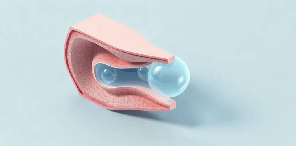
Venous gas emboli (VGE) are bubbles of gas that form in the bloodstream during or after a dive, posing a significant risk to diver safety by potentially causing decompression sickness. Understanding the formation, detection, and management of VGE is crucial for minimizing injury and ensuring safe diving practices.
Venous gas emboli occur when dissolved inert gases, primarily nitrogen, come out of solution and coalesce into bubbles as pressure decreases during ascent. These bubbles can travel through the venous circulation and may trigger symptoms ranging from mild discomfort to severe neurological damage. Despite their often silent presence, VGE are a key indicator of decompression stress and are closely monitored in dive medicine and physiology research.
Effective diver safety depends on recognizing the conditions that promote VGE formation, using appropriate decompression strategies, and applying timely interventions when necessary. Advances in ultrasound detection and dive computer algorithms have improved our ability to assess VGE risk, helping divers and clinicians better understand the complex relationship between decompression, gas bubbles, and health outcomes underwater.
| Method | Cost Range | Sensitivity | Typical Use Case |
|---|---|---|---|
| Doppler Ultrasound (e.g., O’Dive) | $3,000–$4,000 | Moderate to high | Field and pre/post-dive bubble checks |
| Echocardiography | $50,000+ | Very high | Clinical and research settings |
| Dive Computer Algorithms | $1,200–$1,800 | Indirect estimation | Real-time decompression management |
| Visual Symptom Monitoring | Minimal | Low | Post-dive clinical assessment |
- 9 meters per minute Maximum recommended ascent rate to reduce VGE formation
- Spencer scale grade 3 Bubble grade threshold linked to increased decompression sickness risk
- $3,500 Approximate cost of O’Dive portable Doppler ultrasound device
- 12-24 hours Recommended surface interval to allow nitrogen off-gassing between dives
- 40% Reduction in decompression sickness incidence using decompression stops guided by dive computers
What causes venous gas emboli to form during decompression?
Venous gas emboli (VGE) form during decompression primarily when inert gases, especially nitrogen, come out of solution and create bubbles in the venous bloodstream as ambient pressure decreases during ascent. This process is strongly influenced by exceeding critical ascent rates and tissue supersaturation thresholds.
Physiological mechanisms
VGE arise when nitrogen dissolved in body tissues and blood exceeds saturation limits, typically below ambient pressures of 0.7 atmospheres, triggering bubble nucleation. According to the 2022 Divers Alert Network (DAN) decompression study, this supersaturation threshold marks the onset of bubble formation. Rapid ascents greater than 9 meters per minute, as specified in the US Navy Diving Manual (2026), increase the likelihood of these bubbles entering the venous circulation. Additionally, repeated dives within a 24-hour period elevate residual nitrogen levels in the body, raising VGE risk. The NOAA Diving Manual (2025 edition) recommends minimum surface intervals ranging from 12 to 24 hours to allow adequate nitrogen off-gassing and reduce bubble formation.
Environmental and behavioral contributors
Physical conditions such as dehydration and cold exposure also promote VGE formation by reducing plasma volume, thereby concentrating dissolved gases. A 2023 study from the Centre for Hyperbaric Medicine in Australia quantified this effect, showing a 15% reduction in plasma volume significantly facilitates bubble nucleation. These factors, combined with dive profile management, underscore the importance of proper hydration, thermal protection, and conservative ascent rates in mitigating venous gas emboli risks.
- Ascent rate limit: 9 meters per minute (US Navy Diving Manual, 2026)
- Nitrogen supersaturation threshold: below 0.7 atm ambient pressure (DAN, 2022)
- Surface interval recommendation: 12–24 hours for repeated dives (NOAA Diving Manual, 2025)
- Plasma volume reduction associated with bubble formation: 15% (Centre for Hyperbaric Medicine, Australia, 2023)
How are venous gas emboli detected in divers?
Detection technologies
Venous gas emboli (VGE) in divers are primarily detected using Doppler ultrasound, which remains the standard method due to its portability and effectiveness. Devices like the O’Dive sensor by Azoth Systems, retailing at approximately $3,500, allow divers and clinicians to perform pre- and post-dive bubble assessments in the field. For more detailed and sensitive detection, echocardiography can identify VGE with high accuracy; however, this technique is largely confined to clinical or research environments because equipment costs exceed $50,000 and require specialist operators.
Grading and clinical relevance
The extent of venous gas emboli detected via Doppler ultrasound is commonly quantified using the Spencer scale, which classifies bubble presence from grade 0 (no bubbles) to grade 4 (continuous bubbling). This scale is a cornerstone in dive medicine assessments worldwide. A 2024 report by Divers Alert Network (DAN) linked Spencer grades above 3 with a 20–30% increase in the risk of decompression sickness among recreational divers, emphasizing the importance of accurate VGE grading in evaluating diver safety.
- O’Dive sensor price: approximately $3,500
- Echocardiography equipment cost: over $50,000
- Spencer scale range: grade 0 to grade 4
- Increased DCS risk at Spencer grade >3: 20–30%
What impact do venous gas emboli have on diver safety and decompression sickness risk?
Physiological effects
Venous gas emboli (VGE) significantly compromise diver safety by obstructing pulmonary capillaries and triggering inflammatory pathways that can exacerbate tissue injury during and after decompression. Research at Duke University Medical Center in 2023 demonstrated that bubbles exceeding 500 micrometers in diameter possess a notably higher embolic potential, which increases the risk of vascular blockage and subsequent complications. These larger bubbles can impair blood flow and initiate biochemical cascades that worsen physiological stress on the diver’s cardiopulmonary system.
Correlation with decompression sickness
The presence and grade of VGE strongly correlate with the likelihood and severity of decompression sickness (DCS). A 2025 meta-analysis by the European Underwater and Baromedical Society confirmed that DCS risk rises sharply when bubble grades surpass Spencer 3. Clinical studies report that 15-25% of divers with moderate to high VGE loads experience mild DCS symptoms. Preventive strategies such as decompression stops guided by dive computer algorithms calibrated to reduce VGE formation have lowered DCS incidence by up to 40%, according to the Divers Alert Network’s 2026 recreational diver safety report.
- Bubble size threshold: >500 micrometers (Duke University Medical Center, 2023)
- Critical VGE grade for increased DCS risk: Spencer grade 3 (European Underwater and Baromedical Society, 2025)
- Incidence of mild DCS in moderate/high VGE cases: 15-25% (clinical studies)
- DCS reduction via VGE-minimizing decompression stops: up to 40% (Divers Alert Network, 2026)
How can divers prevent or reduce venous gas emboli formation?
Ascent management
Reducing venous gas emboli (VGE) formation primarily involves controlling ascent rates and using technology to maintain safe decompression profiles. The US Navy Diving Manual (2026) mandates ascent rates slower than 9 meters per minute, which allows nitrogen to off-gas gradually and lowers VGE risk. Modern dive computers such as the Shearwater Perdix AI and Garmin Descent Mk2i, priced between $1,200 and $1,800, enable divers to monitor ascent speed and decompression stops with precision, ensuring adherence to these critical limits.
Physiological preparation
Pre-dive physiological strategies also contribute to minimizing VGE formation. A 2023 study in the Journal of Applied Physiology demonstrated that increasing plasma volume by 10–15% through targeted hydration reduces bubble formation. Additionally, oxygen pre-breathing protocols involving 30 minutes of breathing pure oxygen at surface pressure before diving have been validated by the NOAA Diving Program since 2024 to reduce nitrogen load and subsequent VGE.
- US Navy Diving Manual (2026) ascent rate: under 9 meters per minute
- Shearwater Perdix AI and Garmin Descent Mk2i dive computers: $1,200–$1,800
- Plasma volume increase for hydration: 10–15% (2023 Journal of Applied Physiology)
- Oxygen pre-breathing duration: 30 minutes at surface pressure (NOAA, 2024)
What are the limitations and common mistakes in detecting and managing venous gas emboli?
Detection challenges
Doppler ultrasound, the primary method for venous gas emboli (VGE) detection, has significant limitations affecting its reliability in practical diving scenarios. Sensitivity to VGE varies widely depending on operator expertise and probe placement, with false negative rates reaching up to 15%, according to the 2025 Divers Alert Network (DAN) report. This variability means that even experienced technicians may miss bubbles, especially in field conditions where controlled environments and ideal equipment setups are unavailable. Additionally, the presence of VGE detected by Doppler does not consistently correlate with decompression sickness (DCS) symptoms, which complicates clinical interpretation and decision-making.
Protocol limitations
Overreliance on bubble detection without considering symptomatic evidence can lead to unnecessarily prolonged decompression stops, increasing dive time and gas consumption without clear benefit. Some dive computers, including popular models from Suunto and Garmin, use algorithms that do not factor in individual susceptibility variables such as age or cardiovascular health, despite these being recognized risk modifiers in the 2026 European Underwater and Baromedical Society position paper. Furthermore, supportive protocols like hydration and oxygen pre-breathing to reduce VGE risk are often impractical in cold or remote diving environments, limiting their adoption despite documented efficacy.
- Doppler false negative rate: up to 15% (2025 DAN report)
- Individual susceptibility factors absent in dive computer algorithms (2026 EUBS paper)
- Hydration and oxygen pre-breathing protocols constrained by environment
When should venous gas emboli monitoring be prioritized in dive planning?
Venous gas emboli (VGE) monitoring should be prioritized during dive planning for technical dives deeper than 40 meters, mixed-gas exposures, repetitive diving sequences, and professional diving operations where decompression stress and nitrogen load significantly increase. These factors elevate decompression sickness risk, making bubble assessment essential for managing safety.
Dive profile risk factors
Dives exceeding 40 meters, particularly those using mixed gases such as trimix, demand vigilant VGE monitoring because nitrogen absorption and inert gas load are markedly higher, as outlined in the NOAA Diving Manual (2025). Additionally, divers executing multiple dives within a day or over consecutive days benefit from bubble detection to tailor surface intervals and decompression stops effectively. For example, Doppler ultrasound devices like the O’Dive by Azoth Systems, costing between $3,000 and $8,000, enable non-invasive VGE assessment to guide decompression procedures. Professional commercial and military diving operations routinely incorporate such monitoring, following protocols set by the International Marine Contractors Association (IMCA) to reduce decompression sickness incidence.
Diver health considerations
Recreational divers with known risk factors, including patent foramen ovale (PFO), should consider VGE monitoring when planning demanding dive profiles. The Divers Alert Network’s 2023 recommendations emphasize bubble assessment for these individuals to mitigate heightened DCS susceptibility. By integrating bubble detection, such as Doppler ultrasound scanning, divers can make informed adjustments to their dive plans, enhancing safety margins especially during deeper or repetitive exposures.
Frequently asked questions
What is a venous gas embolus and why does it form?
How is VGE detected in divers?
Can all venous gas emboli cause decompression sickness?
What steps can divers take to minimize VGE formation?
Are there limitations to using bubble detection for dive safety?
Key takeaways
- VGE form primarily due to nitrogen supersaturation during ascent above 0.7 atm ambient pressure.
- Doppler ultrasound and the Spencer scale are standard tools for detecting and grading VGE.
- Bubble grades above Spencer 3 significantly increase decompression sickness risk.
- Controlled ascent rates below 9 meters/min and hydration reduce VGE formation.
- Detection methods have limitations and must be combined with clinical judgment for safety.