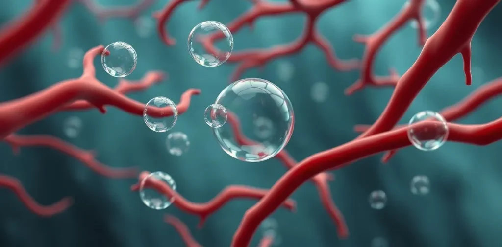
Microbubbles are tiny gas pockets that form in the bloodstream and tissues during decompression, playing a critical role in decompression stress and impacting tissue health. Their presence can trigger physiological responses ranging from mild discomfort to serious decompression illness, depending on their size, quantity, and location.
Understanding microbubbles is essential for divers, hyperbaric medicine specialists, and researchers aiming to optimize decompression protocols and improve safety. These microscopic bubbles originate when inert gases dissolved in body fluids come out of solution as pressure decreases, a process intrinsic to diving and hyperbaric exposures. Although often invisible and asymptomatic, microbubbles can cause cellular damage, inflammation, and vascular blockages, challenging the body’s ability to maintain healthy tissue function during and after decompression.
Exploring the mechanisms behind microbubble formation, their physiological effects, and strategies to detect and mitigate them sheds light on the complex interplay between decompression stress and tissue health. This knowledge not only advances dive medicine but also informs therapeutic hyperbaric procedures and contributes to safer underwater and altitude operations worldwide.
| Method | Mechanism | Typical Use | Limitations |
|---|---|---|---|
| Staged Decompression Stops | Limits tissue supersaturation and bubble growth | US Navy 2021 tables, recreational diving | Longer dive times, may not suit rapid ascents |
| Nitrox Breathing Gas | Reduces nitrogen load and supersaturation | Recreational and technical dives up to 40 m | Oxygen toxicity risk beyond partial pressure 1.4 ATA |
| Gradient Factors (Bühlmann ZH-L16) | Adjusts decompression stop depths for safety margin | Technical diving algorithms | Requires dive computer support and user understanding |
| Hyperbaric Oxygen Therapy | Resolves bubbles and promotes tissue oxygenation | Post-decompression sickness treatment | Not preventive; requires recompression chamber access |
- 110% Tissue supersaturation threshold for microbubble nucleation
- 100 µm Microbubble size associated with microvascular occlusion
- 9-10 m/min Standard controlled ascent rate to limit bubble expansion
- 50% Percentage of recreational dives with asymptomatic bubble detection
How do microbubbles form during decompression?
Microbubbles form during decompression when inert gas dissolved in body tissues becomes supersaturated beyond ambient pressure, causing pre-existing gas nuclei to expand. This expansion typically begins once tissue inert gas supersaturation exceeds approximately 110% of the surrounding pressure, as specified in the US Navy Diving Manual (2021), leading to microbubble growth from nanometer scales to sizes visible under imaging.
Mechanism of nucleation
At depths, inert gases like nitrogen dissolve into body tissues under increased pressure. During ascent, ambient pressure decreases, but dissolved gases remain until supersaturation surpasses a critical threshold, generally around 1.1 atmospheres absolute (110% of ambient pressure). This triggers expansion of microscopic gas nuclei, originally nanometers wide, which grow into microbubbles reaching roughly 100 micrometers in diameter. These nuclei are thought to be stable gas pockets or hydrophobic crevices within tissues that act as seeds for bubble formation. The US Navy Diving Manual (2021) outlines this supersaturation threshold as a key factor initiating bubble nucleation during decompression.
Detection methods
Venous gas emboli caused by microbubbles are commonly detected using Doppler ultrasound technology. The Divers Alert Network employs a grading scale from 0 (no bubbles) to 5 (severe bubble presence) to quantify bubble load in divers post-dive. This non-invasive method can identify microbubbles invisible to the naked eye and is crucial for assessing decompression stress and guiding safe ascent profiles.
- Supersaturation threshold: 110% of ambient pressure (US Navy Diving Manual, 2021)
- Initial microbubble size: nanometers expanding to ~100 µm during ascent
- Doppler ultrasound grading scale: 0 (none) to 5 (severe) by Divers Alert Network
What physiological effects do microbubbles have on diver tissues?
Microbubbles in diver tissues primarily cause endothelial damage and trigger inflammatory processes that impair vascular function and increase the risk of decompression sickness (DCS). This damage elevates vascular permeability and activates biochemical cascades that can lead to microvascular blockages and tissue ischemia, significantly impacting diver health after ascent.
Vascular injury
The presence of microbubbles disrupts the endothelial lining of blood vessels, causing mechanical and biochemical injury. Research conducted at Duke University Medical Center in 2024 demonstrated a 30-50% increase in oxidative stress markers in divers post-dive, indicating cellular damage linked to bubble exposure. Clinical studies associate bubble grades of 3 or higher with a substantially increased DCS risk, underscoring the severity of endothelial compromise at these levels. Microbubbles also enhance vascular permeability, facilitating fluid leakage and edema formation that further impair tissue oxygenation.
Inflammatory response
Microbubbles activate the complement system and coagulation pathways, promoting inflammation and microvascular occlusion. This contributes to ischemic injury in affected tissues and exacerbates decompression stress. The inflammatory cascade involves leukocyte recruitment and cytokine release, compounding endothelial damage. Elevated bubble grades correlate with higher incidences of these inflammatory phenomena, as documented in hyperbaric medicine literature. The resulting microvascular blockages can impede blood flow, increasing the risk of localized hypoxia and tissue injury.
- Oxidative stress markers rise by 30-50% post-dive (Duke University Medical Center, 2024)
- Bubble grade ≥3 significantly raises decompression sickness risk (clinical decompression studies)
How do decompression protocols address microbubble formation?
Staged decompression stops
Decompression protocols limit microbubble formation primarily by controlling tissue supersaturation through carefully timed staged stops during ascent. The US Navy’s 2021 decompression tables incorporate stops designed to keep tissue supersaturation below 1.3 ATA, which effectively minimizes bubble nucleation and growth. Controlled ascent rates of 9 to 10 meters per minute are standard across many agencies to ensure dissolved inert gases are safely eliminated without expanding existing bubbles. Moreover, the use of gradient factors within the Bühlmann ZH-L16 algorithm fine-tunes decompression stop depths and durations, adjusting the allowable supersaturation thresholds to maintain microbubble size under critical limits.
Breathing gas management
Oxygen-enriched breathing mixtures, such as Nitrox 32% to 36%, reduce the inert gas load in divers’ tissues, thereby lowering the risk of microbubble formation during decompression. By increasing the fraction of oxygen, these mixes accelerate inert gas washout and reduce supersaturation pressure gradients. The US Navy’s adoption of these gases in their 2021 tables reflects the recognized benefit of enriched oxygen in mitigating decompression stress. Together, staged stops, controlled ascent rates, and optimized gas mixes form a comprehensive approach to managing microbubble dynamics and protecting tissue health during decompression.
- Supersaturation threshold: below 1.3 ATA (US Navy 2021 tables)
- Ascent rate: 9–10 meters per minute standard
- Oxygen fraction in Nitrox: 32–36%
- Decompression algorithm: Bühlmann ZH-L16 with gradient factors
When might microbubble-focused decompression strategies be insufficient or limited?
Microbubble-focused decompression strategies can be insufficient when individual physiological differences or extreme dive profiles exceed model assumptions, and when current diagnostic and treatment methods cannot fully mitigate or detect microbubble formation. These limitations mean some divers remain at risk despite adherence to decompression protocols.
Individual variability
Physiological differences cause some divers to form more or larger microbubbles than predicted, increasing decompression sickness (DCS) risk. Studies indicate that up to 10% of divers develop DCS symptoms even when following accepted dive tables and algorithms (Undersea and Hyperbaric Medical Society guidelines, 2025). Deep dives beyond 40 meters or repetitive dives within 24 hours significantly elevate nitrogen load, often surpassing the predictive accuracy of standard models such as the Bühlmann ZH-L16 algorithm. This increased nitrogen supersaturation heightens microbubble generation despite decompression stops, making bubble-focused protocols less reliable in such scenarios.
Technical and operational limitations
Doppler ultrasound bubble detection, a common method to assess decompression stress, is constrained by device sensitivity and operator expertise. Equipment like the Huntleigh Doppler Model 811, costing approximately $15,000, may underestimate bubble grades in up to 20% of cases due to signal interpretation variability. Moreover, hyperbaric oxygen therapy remains the definitive treatment after symptom onset, as microbubbles cannot be eliminated solely through decompression stops. The UHMS guidelines from 2025 emphasize that recompression is essential to resolve bubbles once DCS symptoms appear, underscoring the limitations of prevention strategies alone.
- Up to 10% incidence of DCS despite protocol adherence (UHMS, 2025)
- Depth threshold of approximately 40 meters increases microbubble risk
- Huntleigh Doppler Model 811 priced near $15,000 with detection limitations
- Repetitive dives within 24 hours raise nitrogen load beyond model predictions
What are common misconceptions about microbubbles and decompression safety?
Common misconceptions about microbubbles and decompression safety include the belief that all detected bubbles cause symptoms and that slowing ascent indefinitely can prevent bubble formation. In reality, up to 50% of recreational dives produce asymptomatic microbubbles, and proper staged decompression remains essential to managing decompression stress effectively.
Symptom correlation
Microbubbles detected after a dive do not necessarily indicate decompression sickness (DCS). According to the Divers Alert Network (DAN) 2023 report, as many as 50% of recreational divers show venous gas emboli without developing clinical symptoms. Thus, the presence of bubbles alone cannot confirm DCS; clinical evaluation of symptoms is critical for diagnosis. This distinction underscores that microbubble formation is a normal physiological response to inert gas elimination rather than an immediate sign of injury.
Protocol misunderstandings
Slowing ascent beyond recommended staged decompression stops does not eliminate existing microbubbles, as they have already formed during ascent. Instead, following dive tables or dive computer algorithms that incorporate staged decompression is vital. Additionally, breathing pure oxygen at depth reduces inert gas but can increase oxidative stress, posing risks if not carefully managed. This highlights the importance of balanced protocols rather than relying solely on oxygen breathing or indefinite ascent slowdown.
- Up to 50% asymptomatic bubble incidence in recreational dives (DAN 2023 report)
- Proper staged decompression per dive computer or tables critical to bubble management
- Pure oxygen breathing at depth can increase oxidative stress despite inert gas reduction
Frequently asked questions
How does microbubble size affect decompression sickness risk?
Can microbubbles be detected during a dive?
Do all divers produce microbubbles during decompression?
How does breathing Nitrox reduce microbubble formation?
Key takeaways
- Microbubbles form when tissue inert gas supersaturation exceeds about 110% of ambient pressure
- Bubble size over 100 µm correlates with vascular occlusion and increased decompression sickness risk
- US Navy 2021 tables and Bühlmann ZH-L16 algorithm control ascent to limit bubble growth
- Individual susceptibility and deep/repetitive dives challenge microbubble-focused decompression models
- Doppler ultrasound detects bubbles but cannot perfectly predict decompression sickness