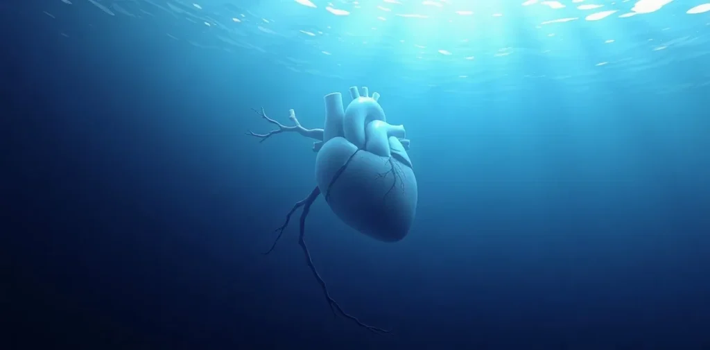
Multiple Brain Lesions in Sport Divers: Are They Associated with a Patent Foramen Ovale? is a 1997 meeting abstract by Knauth and colleagues, published in Undersea & Hyperbaric Medicine. It addresses whether brain lesions observed in sport divers may be associated with a patent foramen ovale, a question relevant to decompression physiology and neurological risk.
What question connects brain lesions and a patent foramen ovale?
The title frames a possible relationship between two features: lesions in the brain of sport divers and a patent foramen ovale, commonly abbreviated PFO. A PFO is a persistent opening between the heart’s upper chambers, left from fetal circulation. In some people it allows blood to pass between chambers without first travelling through the lungs.
The diving-medicine interest is whether this route could affect how gas bubbles or other material move through the circulation. A venous bubble would ordinarily be filtered by the lungs; a pathway that bypasses that filter raises a physiological question about how bubbles might reach the arterial circulation and, potentially, the brain. The title poses an association question, not a diagnosis or an explanation established for every lesion.
Why does a PFO matter in diving physiology?
During and after a dive, inert gas taken up under pressure leaves the body during decompression. Gas bubbles can form in venous blood and tissues, especially when decompression stress is substantial. Most venous bubbles do not cause symptoms, but their movement and effects are central to understanding decompression illness.
A PFO may provide a right-to-left shunt: blood can cross from the right side of the heart to the left, bypassing the lungs. If bubbles cross this route, they can enter arteries, a process known as paradoxical embolisation. This is one proposed mechanism by which a venous decompression event could be associated with neurological effects, although neurological symptoms after diving have multiple possible causes.
What can brain lesions mean in this context?
A brain lesion is a broad imaging description of an area that looks different from surrounding tissue. It is not, by itself, a specific diagnosis or proof of a particular cause. Interpretation depends on the imaging pattern, clinical history and other relevant medical information.
In divers, researchers have considered whether brain-imaging findings might relate to exposure to decompression stress, bubble passage, vascular factors or other causes unrelated to diving. Studying such findings requires care: an association between an anatomical feature and an imaging observation does not establish that one caused the other. The question is important because neurological consequences can affect health and diving fitness, while screening and follow-up decisions must avoid treating uncertain associations as certainty.
- PFO: a persistent opening between the heart’s upper chambers that can permit blood to cross between them.
- Right-to-left shunt: blood passing to the arterial side without going through the lung circulation first.
- Paradoxical embolisation: a possible route by which material from the venous circulation enters the arterial circulation.
- Brain lesion: an imaging finding that needs clinical interpretation and is not a diagnosis on its own.
How does this question fit into diving medicine?
Research on PFO and neurological concerns sits within the wider study of decompression illness, gas-bubble behaviour and individual variation in risk. Diving physicians also distinguish among neurological decompression illness, other dive-related conditions and unrelated neurological disease. The same symptom or scan finding can have more than one explanation, making clinical context essential.
Work in this area examines whether a cardiac shunt is associated with particular clinical or imaging patterns, how a suspected pathway might operate, and what such an association could mean for risk assessment. Those are related but distinct questions. A meeting abstract is part of scientific discussion, while clinical decisions require a broader evaluation than a title or a single research question can provide.
What should divers and clinicians take from the topic?
The subject highlights a plausible connection between cardiovascular anatomy and decompression physiology, not a reason to assume that every PFO is dangerous or that every brain lesion in a diver is dive-related. PFOs are found in people who dive and people who do not, and an anatomical finding alone does not establish an individual’s risk.
Anyone with neurological symptoms after diving should seek prompt medical assessment, and divers concerned about a PFO or an imaging finding should consult a physician experienced in diving medicine. Decisions about investigation, treatment or future diving need individual assessment; general information cannot determine fitness to dive.
Frequently asked questions
What is a patent foramen ovale?
How could a PFO be relevant to decompression?
Does a brain lesion on imaging identify its cause?
Citation details
- Title: Multiple Brain Lesions in Sport Divers: Are They Associated with a Patent Foramen Ovale?
- Authors: Knauth, M; Ries, S; Forsting, M; Daffertshofer, M; Hennerici, M; Sartor, K
- Year: 1997
- Published in: Undersea & Hyperbaric Medicine 1997
- Record type: Meeting abstract
- Repository record: Rubicon Research Repository, handle 123456789/374
This page is an original summary written by the Rubicon editors from the publication’s bibliographic record. It does not reproduce the paper, its abstract or its data; consult the publication itself for its methods and findings.
Related reading on Rubicon
- Scientific Diver vs. Recreational Diver
- Why Divers Get Cold Faster Underwater
- Scuba Diving Equipment: Essential Gear for Every Diver
- Effects of Cold Water Immersion on Diver Metabolism
- More on Dive Medicine
- Browse the Research Library
