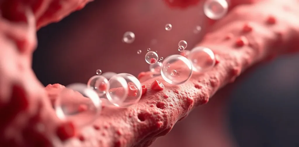
What causes inert gas bubbles to form during decompression?
Inert gas bubbles form during decompression when dissolved gases, primarily nitrogen or helium absorbed at depth, come out of solution as ambient pressure decreases. This process occurs because tissues become supersaturated—meaning the partial pressure of dissolved gas exceeds the ambient pressure—leading to nucleation and growth of gas bubbles. The fundamental trigger is a reduction in ambient pressure faster than gas can be eliminated via respiration, causing free gas pockets to develop.
Bubble formation starts at microscopic nuclei, which are pre-existing gas pockets or hydrophobic crevices in tissues. These serve as sites where dissolved gas can coalesce. Supersaturation thresholds vary by tissue type but often begin around 1.5 to 2 atmospheres of inert gas supersaturation. This mechanism is the physiological basis for decompression sickness (DCS), making controlled ascent profiles essential.
How does tissue type affect bubble formation and risk?
Tissue composition and perfusion significantly influence bubble formation. Fast-perfused tissues like blood and lungs equilibrate gases rapidly, while slow tissues such as fat absorb and release inert gases more slowly, affecting supersaturation dynamics.
Tissue-specific factors
- Blood and lungs: Rapid gas exchange; bubble formation less likely due to quick inert gas clearance.
- Fat tissue: High solubility for nitrogen; slow washout increases residual gas tension.
- Muscle and connective tissue: Intermediate perfusion; moderate risk for bubble nucleation.
These differences require decompression algorithms like Bühlmann ZHL-16C to use multiple tissue compartments with varying half-times from 4 to 635 minutes to model bubble risk accurately. The choice of these compartments affects dive computer safety settings.
What role do supersaturation gradients and ascent rates play?
Supersaturation gradients—the difference between tissue inert gas partial pressure and ambient pressure—drive bubble formation. A steep gradient increases the likelihood of bubble nuclei expanding into harmful bubbles.
Ascent rate is a critical factor; ascending too quickly causes excessive supersaturation. Standards such as those from NOAA recommend ascent rates of no more than 9 meters per minute to manage this risk. In technical diving, slower rates or decompression stops are employed to control gradients and allow inert gas elimination.
Ascent protocols and bubble control
- Standard recreational ascent: 9 m/min maximum with safety stop at 5 meters for 3-5 minutes.
- Technical decompression: Incorporates multiple stops, often starting deeper, with stop durations tailored to tissue compartments.
- Deep stops: Used to reduce bubble growth in fast tissues, though their efficacy remains under study.
How do microbubbles and venous gas emboli impact diver safety?
Microbubbles are microscopic gas bubbles that form in venous blood during decompression; when detected by Doppler ultrasound, they are termed venous gas emboli (VGE). VGE presence correlates with increased risk of DCS but not all divers with VGE develop symptoms.
Monitoring VGE incidence helps assess decompression stress. Studies have found that bubble grades measured by the Spencer scale or Kisman-Masurel scale correlate with DCS risk. For example, a high bubble grade (>3 on the Spencer scale) corresponds to a significantly elevated risk.
Bubble monitoring methods
- Doppler ultrasound: Noninvasive detection of VGE in subclavian or precordial veins.
- Echocardiography: More sensitive, can detect arterialized bubbles crossing a patent foramen ovale (PFO).
- Bubble grading scales: Standardized systems for quantifying bubble load and risk assessment.
| Scale | Bubble Grade Range | Risk Correlation |
|---|---|---|
| Spencer Scale | 0–5 | Grades ≥3 indicate moderate to high DCS risk |
| Kisman-Masurel Scale | 0–IV | Grades III–IV correlate with significant bubble load |
What physiological mechanisms influence bubble growth and elimination?
Once formed, bubble growth depends on factors such as tissue gas tension, ambient pressure, and surface tension at the bubble interface. The body eliminates bubbles primarily by diffusion of gas into blood and exhalation via lungs.
Endothelial responses and immune activation also modulate bubble effects; bubbles can trigger inflammation, platelet aggregation, and endothelial damage contributing to DCS pathology. The body’s ability to control bubble size through these mechanisms is vital.
Factors affecting bubble dynamics
- Gas diffusion: Driven by partial pressure gradients; oxygen breathing accelerates inert gas washout.
- Bubble surface tension: Smaller bubbles have higher internal pressure, influencing stability.
- Physiological response: Activation of nitric oxide release can reduce bubble adhesion and vascular injury.
How do decompression models incorporate bubble formation science?
Modern decompression algorithms integrate bubble physics to improve diver safety. Bubble models such as the Varying Permeability Model (VPM) and Reduced Gradient Bubble Model (RGBM) predict bubble formation and growth by simulating gas dynamics in tissues.
These models differ from traditional dissolved gas models by explicitly accounting for bubble nuclei behavior and propose ascent schedules that limit bubble growth rather than just tissue supersaturation.
Popular decompression algorithms
- Bühlmann ZHL-16C: Widely used, models 16 compartments with fixed supersaturation limits.
- Varying Permeability Model (VPM): Focuses on controlling bubble nuclei size and number; used by companies like Shearwater Research.
- Reduced Gradient Bubble Model (RGBM): Emphasizes conservative profiles with bubble management; implemented in the Suunto EON Core dive computer.
| Algorithm | Approach | Key Implementation |
|---|---|---|
| Bühlmann ZHL-16C | Dissolved gas supersaturation limits | Used in Garmin Descent Mk2i dive computer |
| VPM | Bubble nuclei control | Shearwater Perdix AI dive computer |
| RGBM | Bubble growth reduction with conservative stops | Suunto EON Core dive computer |
Frequently asked questions
Why do bubbles form more readily during rapid ascents?
Can bubbles form without symptoms of decompression sickness?
How do decompression stops reduce bubble formation?
Are some tissues more susceptible to bubble formation?
Key takeaways
- Bubble formation occurs when tissue inert gas supersaturation exceeds ambient pressure during decompression.
- Tissue perfusion rates and gas solubility affect bubble nucleation and growth.
- Controlled ascent rates and decompression stops mitigate dangerous supersaturation gradients.
- Venous gas emboli detection helps evaluate decompression stress and DCS risk.
- Modern decompression algorithms incorporate bubble physics to optimize diver safety.
Conclusion
Understanding the physiological mechanisms behind inert gas bubble formation during decompression is crucial for enhancing diver safety and preventing decompression sickness. Supersaturation, tissue-specific gas dynamics, and controlled ascent protocols collectively determine bubble risk. Advances in bubble modeling and monitoring continue to refine decompression strategies, underscoring the importance of integrating bubble science into diving practices.