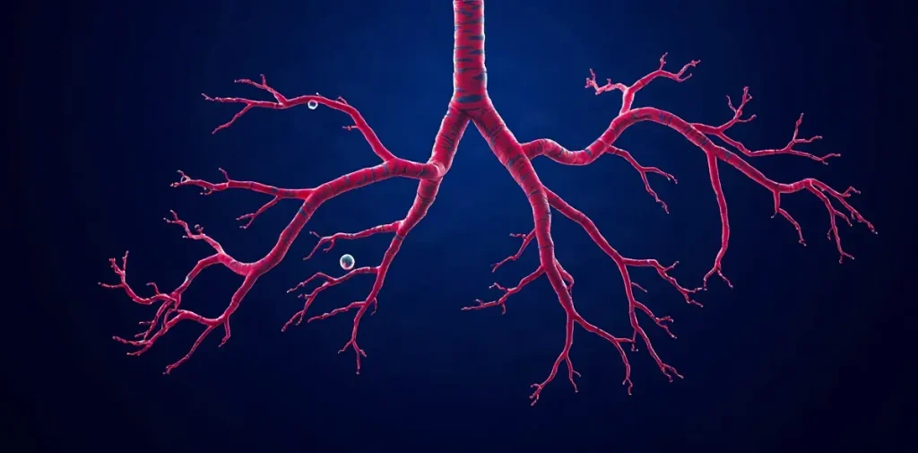
Bubble formation in the pulmonary vasculature concerns how gas bubbles may arise within the blood vessels of the lungs under changing pressure conditions. The question matters because the lungs are both a site where circulating bubbles encounter a dense capillary network and a key part of the body’s response to decompression.
Why focus on the lungs and blood vessels?
During a dive, increased ambient pressure raises the amount of inert gas that dissolves in body tissues. As pressure falls during ascent, dissolved gas can come out of solution and form bubbles. Those bubbles may circulate in venous blood toward the right side of the heart and then the pulmonary arteries, where the lung’s branching vessels filter and exchange gases.
The pulmonary circulation is not simply a passive sieve. Bubbles can deform, divide, lodge temporarily or pass through, depending on their size, flow and the properties of the vessel. The lung also has a large surface area for gas exchange, so bubble behaviour there links decompression physiology to respiratory and cardiovascular function.
Studying bubble formation in this setting asks a distinct question from measuring how many bubbles appear after a dive. It concerns the physical and physiological conditions under which gas can begin to form in or near pulmonary vessels, and how local pressure differences might contribute.
What does a pressure gradient have to do with bubbles?
A pressure gradient is a change in pressure between neighbouring regions. In the pulmonary circulation, blood moves through vessels that branch repeatedly and narrow toward the capillaries; pressure and flow vary across this network. A local pressure difference may affect whether dissolved gas remains in solution, grows at a pre-existing gas nucleus or forms a bubble.
Bubble formation is not governed by pressure alone. Gas tension, the blood and vessel environment, flow, and the presence of microscopic nuclei can all influence whether a bubble starts and whether it grows. A nucleus is a tiny gas-containing site that can make bubble growth possible under conditions where a completely new gas–liquid boundary would be difficult to create.
The term «minimum delta P» points to a threshold question: what pressure difference might be required for bubble formation in this setting? A threshold is not automatically a universal safety limit. It depends on the experimental conditions and on how the pressure difference and bubble formation are defined and measured.
How do researchers investigate the question?
Work on bubble formation combines concepts from gas physics, fluid mechanics and physiology. Researchers can examine how pressure changes affect gas dissolved in a liquid, how flow through vessels changes local conditions, and what happens when bubbles encounter narrow channels. Experimental models can isolate parts of the process, while physiological studies address the complexity of living circulation.
Each approach has trade-offs. A simplified model makes pressure, flow or gas content easier to control, but may not reproduce the full behaviour of blood vessels and tissues. Measurements in a living system are more physiologically relevant, yet it can be harder to separate bubble formation from transport, growth and clearance.
- Initiation: whether a bubble begins to form, rather than merely arriving from elsewhere.
- Growth: how dissolved gas may add to an existing bubble.
- Transport: how blood flow carries bubbles through the pulmonary vessels.
- Detection: whether an observed signal indicates a bubble, and where it is located.
These distinctions matter because a detected bubble does not by itself reveal where it formed, what caused it, or what effect it will have.
Why does this matter for decompression and clinical care?
Decompression illness involves gas bubbles and their effects, but the presence of bubbles does not by itself establish that a diver is ill. Bubble number, location, movement and interaction with tissues all matter, as do the diver’s symptoms and clinical condition. Understanding pulmonary bubble behaviour helps researchers frame how bubbles are filtered, redistributed or associated with physiological stress.
The subject also connects to broader decompression research: models of inert-gas uptake and elimination, studies of bubble nuclei, and investigations of how ascent profiles affect bubble formation. These are related but not interchangeable questions. A mechanism observed in a controlled model cannot, on its own, determine a safe ascent profile for every diver or exposure.
For divers and scientific teams, this work contributes to the reasoning behind decompression procedures and medical assessment rather than offering a stand-alone rule. Suspected decompression illness requires prompt assessment by a diving-medicine physician or emergency service; an individual dive plan should not be changed on the basis of a general explanation.
What can a threshold tell us—and what can it not?
A measured threshold can help identify conditions under which bubble formation becomes more likely in a specified system. Its usefulness depends on whether the system resembles the physiology of interest, how pressure and bubbles are measured, and whether the result can be reproduced under other conditions.
Even a well-defined threshold would describe one part of a larger process. Divers differ in exposure and physiology, and decompression involves changing gas tensions across multiple tissues and circulation pathways. The practical question is therefore not only when a bubble can form, but how formation, growth and transport combine with the diver’s condition.
Frequently asked questions
Does finding bubbles mean a diver has decompression illness?
Is a pressure threshold the same as a safe diving limit?
Why study bubbles in pulmonary vessels rather than only in tissues?
An original research-library explainer written by the Rubicon editors. It is not a summary of a specific publication.