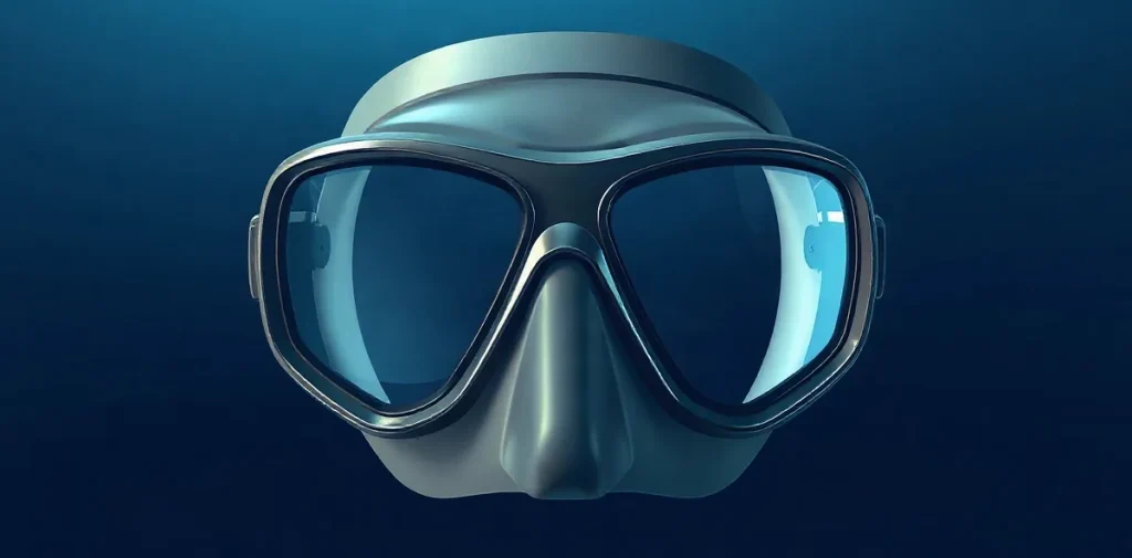
Orbital hemorrhage after face-mask barotrauma concerns bleeding within or around the eye socket when pressure changes during a dive injure delicate tissues. It connects a familiar equipment problem—the mask squeeze—to eye anatomy, pressure physiology and clinical assessment. Understanding that link helps divers and clinicians distinguish a local pressure injury from other causes of eye symptoms.
How can a face mask injure the tissues around the eye?
A diver’s mask encloses air in front of the eyes and nose. As the diver descends, surrounding water pressure rises; if the gas space is not equalised, pressure inside the mask becomes lower than the pressure outside. The resulting pressure difference can draw soft tissue toward the mask and create a squeeze.
The eye socket contains the globe, muscles, vessels, nerves and cushioning tissue. Small blood vessels can be vulnerable to mechanical stress, while the eyelids and tissues around the orbit may also show bruising or swelling. The term orbital hemorrhage describes bleeding in the region of the orbit; it is distinct from a surface bruise, though symptoms can overlap.
Equalising the mask means adding gas through the nose as ambient pressure increases. A leak, poor fit or delayed equalisation can contribute to pressure imbalance, but the precise circumstances vary from dive to dive.
What does the pressure injury involve?
Barotrauma is tissue injury caused by a pressure difference across an air-containing space or a body surface. In mask squeeze, the relevant pressure imbalance acts locally around the face and eyes, rather than arising from dissolved inert gas leaving the body during decompression. That distinction matters: the mechanism and clinical assessment are not the same as decompression sickness.
Bleeding may occur when vessels are stretched or compressed, but symptoms alone do not establish where bleeding has occurred or how serious it is. Redness, bruising, swelling, pain, altered vision or restricted eye movement call for attention because different structures can be involved.
Eye complaints after diving also have other possible causes, including unrelated eye disease and injuries from impact or equipment. A clinician considers the dive history alongside examination findings, rather than treating every red eye after a dive as the same condition.
How do clinicians assess an eye injury after diving?
Assessment begins with the timing and pattern of symptoms, the dive profile, mask equalisation and any other pressure-related problems. Clinicians examine vision and the eye and surrounding tissues; further evaluation depends on the suspected location and severity of injury. The aim is to identify whether the problem is limited to superficial tissue or involves structures that need urgent care.
- Pressure mechanism: whether the mask air space was equalised during descent.
- Symptoms: when they began and whether vision, movement, pain or swelling has changed.
- Examination: the condition of the eye, eyelids and tissues around the orbit.
- Other explanations: whether another eye condition or injury could account for the findings.
New visual disturbance, significant pain or rapidly increasing swelling should be treated as a reason for prompt medical assessment. For suspected dive-related eye injury, consult a diving-medicine physician or an appropriate eye-care clinician; an online description cannot determine the diagnosis.
Why does this matter beyond one mask squeeze?
Diving medicine studies pressure injuries across several settings: ears and sinuses, lungs, teeth, the gastrointestinal tract and spaces created by equipment. Mask squeeze is a useful reminder that a pressure problem can affect a small, anatomically complex area even when the diver has no obvious difficulty elsewhere.
Case-based clinical work in this area helps describe how an uncommon or easily overlooked injury presents and how it should be distinguished from other conditions. It also links equipment use to physiology: mask fit and equalisation technique shape the pressure exposure, while anatomy and individual health influence the consequences.
For divers, the practical lesson is to equalise the mask gently during descent and stop if equalisation is not working. Do not continue descending through facial pain or pressure; a persistent eye symptom after a dive deserves clinical review before returning to diving.
Frequently asked questions
What is mask squeeze?
Is orbital hemorrhage the same as a decompression illness?
When should a diver seek medical assessment?
An original research-library explainer written by the Rubicon editors. It is not a summary of a specific publication.
Related reading on Rubicon
- Scuba Equipment: Stop Your Mask Fogging
- Full-Face vs Traditional Dive Masks: Usability and Safety
- Comparative Review of Dive Masks for Wide Field of Vision
- Common Causes of Mask Fogging and How to Prevent It
- Otic and Sinus Barotrauma in Hyperbaric Chambers
- Diver Breathing: Mouthpiece vs Full-Face Mask
- More on Dive Medicine
- Browse the Research Library