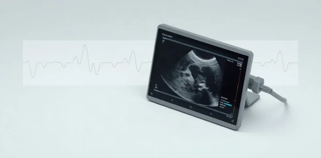
Transcranial Doppler is an ultrasound method for monitoring blood flow through major vessels in the brain. In diving medicine, it offers a way to investigate how circulating gas bubbles and changes in pressure may relate to cerebral circulation—questions relevant to decompression, neurological symptoms and the interpretation of physiological measurements.
What does transcranial Doppler measure?
Ultrasound waves can pass through thin regions of the skull, allowing a probe to detect blood moving in certain intracranial arteries. The instrument records the motion of blood cells as Doppler shifts: changes in the returning sound frequency caused by movement. This provides a non-invasive view of flow in selected vessels, rather than an image of the whole brain or a direct measure of brain function.
The signal is typically described through features such as flow direction and velocity over time. These measurements depend on the vessel being sampled, the probe position and the conditions during monitoring. A Doppler trace is therefore a physiological measurement to interpret in context, not a diagnosis by itself.
Why is blood-flow monitoring relevant to divers?
During and after a dive, pressure changes affect the gases dissolved in the body. Decompression can produce venous gas bubbles, which may travel through the circulation; their presence and movement are of interest because gas emboli can be associated with decompression illness. Transcranial Doppler can be used in research to detect signals consistent with bubbles reaching the cerebral arterial circulation.
That connection is not a simple test of whether a diver is safe or injured. Bubble signals, cerebral blood flow and clinical symptoms are related but distinct observations. A person’s symptoms require medical assessment, and a monitoring result should not be treated as a stand-alone reason to diagnose or exclude decompression illness.
How do researchers use the method?
Studies may monitor blood flow or bubble signals during defined conditions, including before and after diving or during decompression-related investigations. Researchers need to account for the timing of measurements, the dive profile, the probe location and the quality of the acoustic signal. These factors affect what can be compared across observations.
Transcranial Doppler belongs to a wider effort to connect measurable physiological events with the mechanisms and clinical presentation of decompression illness. It can complement other observations, such as symptoms and measurements of bubbles in the venous circulation, but each method answers a different question. The value lies in combining appropriate measurements, not treating one instrument as a complete account of risk.
- Flow velocity: the speed of blood movement in the vessel being sampled, interpreted with the measurement conditions.
- Embolic signals: brief Doppler signals that may be consistent with moving gas bubbles or other embolic material.
- Monitoring: repeated observation over time, useful for studying changes that a single measurement could miss.
- Clinical context: dive exposure, symptoms and examination findings needed to interpret a physiological trace.
What are the limits of a Doppler signal?
The method samples selected vessels through acoustic windows in the skull; it does not map every cerebral vessel or show all brain tissue. Signal quality and interpretation can vary with probe placement and the conditions of recording. A detected signal also needs careful classification: the instrument records sound changes, while the investigator determines what they may represent.
Most importantly, monitoring does not establish a direct cause-and-effect link between a bubble signal and neurological injury. Research must distinguish a marker of exposure or circulation from a clinically meaningful outcome. For divers and clinicians, that means Doppler findings are best understood alongside the dive history, examination and other relevant tests.
How does this research inform diving practice?
For diving physicians and hyperbaric clinicians, cerebral Doppler monitoring helps frame questions about decompression physiology: when signals occur, how they change, and how they relate to other measures. The method can also support research into individual variation and the processes that connect pressure exposure, gas bubbles and neurological effects. Those questions matter even when a measurement is not itself a clinical decision rule.
For divers, the practical distinction is between a research tool and an assessment of personal fitness or symptoms. New neurological symptoms after diving need prompt evaluation by a diving-medicine physician or other appropriate medical professional. A Doppler measurement cannot replace that evaluation.
Frequently asked questions
Does transcranial Doppler show the brain?
Can a bubble signal diagnose decompression illness?
When should a diver seek medical assessment?
An original research-library explainer written by the Rubicon editors. It is not a summary of a specific publication.
Related reading on Rubicon
- The Diving Reflex: What Happens Underwater
- Scuba Diving Fins: Split vs. Paddle Designs
- Scuba Diving Regulators: Balanced vs. Unbalanced
- Scuba Diving Buddy Checks: Catch Problems Early
- Transcranial Doppler Monitoring in Diving Medicine
- Thesis Research in Diving and Hyperbaric Medicine
- More on Dive Medicine
- Browse the Research Library